Welcome to the forum, and to the wonderful world of microscopy.
TL;DR The easiest would be to use their “Mobile Phone Stand” at the eye piece (more details below). EDIT: I was wrong, we wouldn’t know precisely what the magnification is with the mobile phone but I hope some information in my reply is still relevant.

If you have mounted your camera onto the camera port (orange arrow in the picture below). Then, it is normal that you are limited to 4, 10, 40, and 100X.

In an infinity-corrected microscope, and I believe that this is what we are dealing with here, an image of the sample is created by the objective lens and a second lens that is called a tube lens. The magnification of that image is given by the ratio tube lens focal length over objective lens focal length. If you look at the lenses you find several numbers inscribed, for the yellow objective in the screenshot below, you have 10, this is 10X magnification, and then 0.25 (separated by a slash), this is the numerical aperture and gives you an indication on the resolution (among other things). In the second line you see 160, this is the tube lens focal length in mm, 160 mm. Lastly, you have 0.17 (separated by a slash), this is the expected cover glass thickness in mm, or 170um.
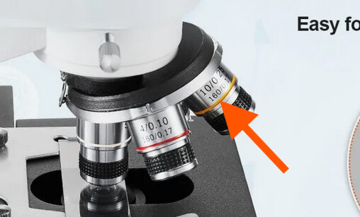
So, the tube lens has a focal length of 160 mm, and if it is a 10X, the objective lens must have a focal length of 16 mm (160 / 16 = 10X) and when you use the camera port, this is what the camera will experience. The camera is located exactly at the image plane.
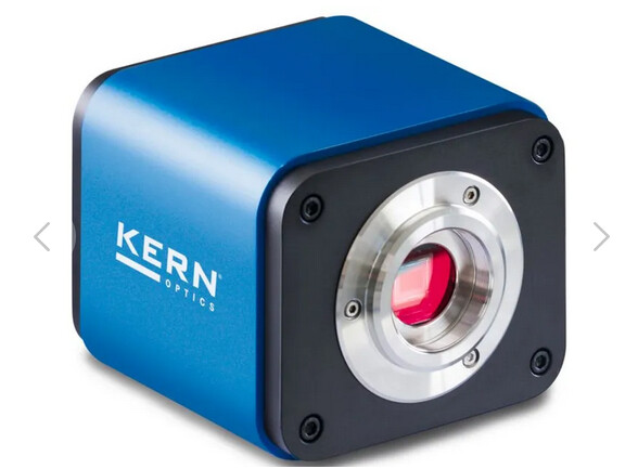
You could also see this image with your eye but you have to think of your eye as being a lens plus a detector. The consequence is that you can hardly focus on objects close to your eyes. This means you have to be a reasonable distance away for an object to see it in focus. Since this image is quite small you’ll have a hard time resolving the details in the image. All of this to say, that is why you need an eyepiece to get a good image with your eyes. But don’t forget that your eye has this additional lens and you cannot simply put your camera in front of the eyepiece. I should be careful here, what I call camera is the Lazmin that you are referring to (although I am not sure I found the exact right one on the internet).
As you can see in the screenshot below, it is only a camera sensor and if you use it alone you will hardly see an image. That is until you place it in the image plane of the microscope.
One thing you could do is use the Mobile Phone Stand that they advertise to take advantage of the additional magnification offered by the eyepiece. This will work because your cellphone camera is a sensor AND a lens, just like your eye (in a way…). If you are interested, I made some basic simulation for a question in image.sc here. EDIT: although as we discussed in the image.sc thread, the final magnification will depend on the cellphone camera lens focal length (and you might not know it), therefore you probably won’t achieve 1000X.
There would be other (more costly and complicated) solutions, but I think it is the most straightforward. Let me us know if you need such a different solution (I feel my answer is already really long…).
One last thing to consider regarding magnification. There’s a good reason why you won’t easily find an objective lens above 100X. In short, diffraction will limit the resolution to about 200nm with a good lens (and oil…), and the pixels are also (de-)magnified in your image. So, a typical 6.5um pixel will have a size of 6.5/100 = 65nm in your image, this is reasonable and satisfy the sampling theorem. With a magnification of 1000X, the pixel will become 6.5nm, and the resolution will still be limited to 200nm by diffraction. Therefore, a lot of pixels will have the same information and we call that empty magnification. I feel like I keep advertising myself but I wrote something about this in a Physics Stack Exchange question. Maybe you will find it interesting too.
Anyhow, let us know if this was useful, and if you have any other question.
Happy imaging ![]()
Omni