Hello, I am new to the forum and a beginner in microscopy. I bought a VEVOR Composite Triocular Microscope 40X-5000X Biological Microscope and a Lazmin112 Digital Microscope Camera, 8.3 MP 1/1.8 CMOS Sensor 23.2 30-mm. the combinations are as follow: 4x10, 4x25, 10x40, 10x100, 25x100 and oil. But if i want to see through the camera, i only have 4x, 10x, 40x, 100x. So my question is: If I want to enhance something to 1000x, how can I do that through the camera lens? Sorry for the bad description, not familiar with the names of all the components.
Welcome to the forum, and to the wonderful world of microscopy.
TL;DR The easiest would be to use their “Mobile Phone Stand” at the eye piece (more details below). EDIT: I was wrong, we wouldn’t know precisely what the magnification is with the mobile phone but I hope some information in my reply is still relevant.

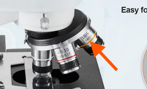
If you have mounted your camera onto the camera port (orange arrow in the picture below). Then, it is normal that you are limited to 4, 10, 40, and 100X.

In an infinity-corrected microscope, and I believe that this is what we are dealing with here, an image of the sample is created by the objective lens and a second lens that is called a tube lens. The magnification of that image is given by the ratio tube lens focal length over objective lens focal length. If you look at the lenses you find several numbers inscribed, for the yellow objective in the screenshot below, you have 10, this is 10X magnification, and then 0.25 (separated by a slash), this is the numerical aperture and gives you an indication on the resolution (among other things). In the second line you see 160, this is the tube lens focal length in mm, 160 mm. Lastly, you have 0.17 (separated by a slash), this is the expected cover glass thickness in mm, or 170um.
So, the tube lens has a focal length of 160 mm, and if it is a 10X, the objective lens must have a focal length of 16 mm (160 / 16 = 10X) and when you use the camera port, this is what the camera will experience. The camera is located exactly at the image plane.
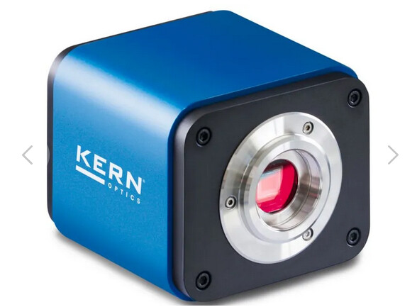
You could also see this image with your eye but you have to think of your eye as being a lens plus a detector. The consequence is that you can hardly focus on objects close to your eyes. This means you have to be a reasonable distance away for an object to see it in focus. Since this image is quite small you’ll have a hard time resolving the details in the image. All of this to say, that is why you need an eyepiece to get a good image with your eyes. But don’t forget that your eye has this additional lens and you cannot simply put your camera in front of the eyepiece. I should be careful here, what I call camera is the Lazmin that you are referring to (although I am not sure I found the exact right one on the internet).
As you can see in the screenshot below, it is only a camera sensor and if you use it alone you will hardly see an image. That is until you place it in the image plane of the microscope.
One thing you could do is use the Mobile Phone Stand that they advertise to take advantage of the additional magnification offered by the eyepiece. This will work because your cellphone camera is a sensor AND a lens, just like your eye (in a way…). If you are interested, I made some basic simulation for a question in image.sc here. EDIT: although as we discussed in the image.sc thread, the final magnification will depend on the cellphone camera lens focal length (and you might not know it), therefore you probably won’t achieve 1000X.
There would be other (more costly and complicated) solutions, but I think it is the most straightforward. Let me us know if you need such a different solution (I feel my answer is already really long…).
One last thing to consider regarding magnification. There’s a good reason why you won’t easily find an objective lens above 100X. In short, diffraction will limit the resolution to about 200nm with a good lens (and oil…), and the pixels are also (de-)magnified in your image. So, a typical 6.5um pixel will have a size of 6.5/100 = 65nm in your image, this is reasonable and satisfy the sampling theorem. With a magnification of 1000X, the pixel will become 6.5nm, and the resolution will still be limited to 200nm by diffraction. Therefore, a lot of pixels will have the same information and we call that empty magnification. I feel like I keep advertising myself but I wrote something about this in a Physics Stack Exchange question. Maybe you will find it interesting too.
Anyhow, let us know if this was useful, and if you have any other question.
Happy imaging ![]()
Omni
Thank you for a wonderfully detailed reply. I’ll have to read it several times to fully understand it.
Here are the product links:
Camera: https://www.amazon.se/-/en/dp/B0FG1B4DKP?ref=ppx_yo2ov_dt_b_fed_asin_title
1 Like
Apologies, I have written a bit too fast. Because the magnification you get from the eye piece is also dependent on the cellphone camera lens focal length (and it is generally not obvious to find this information. I have edited my answer accordingly.
In the end, I am not so sure this is the best solution anymore. I might delete my answer, I don’t know if it is relevant anymore… Sorry for that really, got carried away.
Do you need exactly 1000X magnification?
Take care,
Omni
Thank you again for such pedagogic answers. I am begining to understand. Of course i do not need 1000x mag. We just hope to see some tardigrades, maybe a few face mites ![]() . I Will try the mobile phone and let you know
. I Will try the mobile phone and let you know
1 Like
I was thinking about this overnight. The average focal length of the human eye is about 17mm according to this article. However, cellphone camera lenses are probably more in the range of 4-5mm. This is a factor of approximately 4 times shorter on the focal length and should translate roughly to a factor 1/4 on the eyepiece magnification, assuming the camera lens focal length is still larger than the eyepiece focal length. So if you use 100X times 10X to get 1000X, with the cell phone it might be more like 100X times 10X times 1/4 or 250X. This also assumes that my picture of this microscope arrangement is correct, which at this point is not given. I am starting to thing that you are more like in a compound microscope arrangement on the eyepiece light path, and perhaps still infinity corrected on the camera port light path. There are two things that might help.
- Try to image a ruler, or something of known physical size, use Fiji or any other tool to calculate the known thing size in pixels. Then divide the length of your ruler by the size in pixels. For example, you measure a 1-mm graduation on a ruler, and the distance between the two lines are 1000 pixels (totally random numbers), this gives you 1/1000 = 0.001mm = 1um pixel size (in the image). Your camera has pixels that are 2um in size (physical size). With those two values, you know that the magnification is 2um / 1um = 2X
- Try to get a feeling for the eyepiece focal length. The way I do that is I go under a ceiling lamp and hold the eyepiece vertically above a flat surface, then I approach the eyepiece towards the flat surface until I see an image of the ceiling lamp on the flat surface. I made a short video for you using a Leica stereo microscope 10X eyepiece (10450630). The distance between the eyepiece and the flat surface when an image of the ceiling lamp is formed is roughly in the range of the focal length
Maybe @P_Tadrous can correct my many mistakes!!!
Take care,
Omni
@Omnistic When I posted my question, i never imagined I would receive such detailed and relevant answers. Even if i repeat myself, thank you for sharing your knowledge. With that said, I think your video clarified my question as to why the pictures “suck”. I still have the possibility of returning the microscope to Amazon(no questions asked). The Vevor microscope has also a feel of low quality wheel adjustment, i’d rather return it. Question that remains is what should I get instead. I won’t say that “money is no object” but let’s say maximum of 700 euros. Would this one be a better choice? https://www.amazon.se/-/en/dp/B0FQTT4K55/ref=sr_1_103?dib=eyJ2IjoiMSJ9.r7EHGmMP2SZSEkSZWLuOf1sZyeATmuRYtCu78LtthbrmKsjBS6YHbg4nQE3UB1BzTn2IXLHS2MTW6bz_SjD3P54Oxo9CGghT5MfEcTrt9g4y4hvmcmmrRG_BEjiXyYMqRAXyweh-Z6JJzHhvwVQGzhLhAzH0qpNLuticOyB1gGY1BUIkRll-Th0HVwur8ncEdB5LrskYOuErYmUcsmO0G29Nn9JQ2H8BbjmQRo-sWW3ub4JCIKYW3pH7mR56ckQgOXuo8jYmBNO9r4WlOCbAtWIzwUfRQPM3o2cyCr9Wa0Y.5eJ1TnIO0pYF3TiShR98sk4yI3__awlZAqXHOJ4Yj-Y&dib_tag=se&qid=1758787689&s=industrial&sr=1-103&xpid=iTBYEe4IrylIu
Feel free to make a suggestion.
/Mike
@micke my pleasure really. Do you have pictures that you can share? I’m thinking it should be possible to see tardigrades with this scope (although I don’t know the quality of the build as you said). How are you preparing the sample? Are you using glass cover slips with 170um thickness (those are generally labeled # 1.5)? And what is your mounting medium?
Setting aside the eyepiece magnification. Let’s say you want to image a tardigrade. The length of a tardigrade is 0.05 to 0.5mm according to Wkikipedia. From this size we can draw a few extreme cases to get a feeling for what device you might need.
One case is that you want the biggest tardigrade to be within your field of view. In this case, your sensor has a size 3840 x 2160 in pixels and one pixel is 2um x 2um, so the sensor size in mm is 7.68mm x 4.32mm. Let’s focus on the smallest dimension of your sensor or 4.32mm. Already here you see that if you were using 10X magnification, the large 0.5mm tardigrade would appear with a size of 0.5 * 10 = 5mm on your sensor. Depending on the orientation, it might be cropped already (assuming there is no vignetting). In this image, the pixels will have an apparent size of 2um divided by the magnification (10X) or 0.2um or 200nm. In this case, it is likely that the resolution is not limited by the pixel size. Instead, the resolution is probably limited by diffraction. To get an idea of the diffraction-limited resolution you can use the Abbe formula, wavelength divided by twice the numerical aperture (NA). There are some assumption behind this formula and in your case I would be wary of your illumination NA, but let’s just use this formula anyway to get an idea. The 10X of the Vevor scope seems to be 0.25NA, if you take a wavelength in the center of the visible spectrum like 0.55um. This gives you a theoretical diffraction-limited resolution of 0.55 / (2 * 0.25) = 1.1um. Since it is more than double the pixel size (in the image) of 0.2um, you are satisfying the sampling criterion. Once again, in practice, it doesn’t hurt to go a bit beyond with the sampling (meaning having even smaller pixels, something in the range diffraction-limited resolution divided by 3 or 4). But if the pixels become too small, and the resolution is anyway clearly limited by diffraction, then you get into empty magnification.
Another interesting case is to look at what would happen if you were using the 100X oil, which has a NA of 1.25. The diffraction-limited resolution (at 0.55um) is 0.22um or 220nm. At 100X your pixels also have a size of 2um / 100 = 0.02um or 20nm. The pixels are now roughly ten times smaller than your resolution and I would argue that we are already airing on the side of empty magnification. So, the resolution overall still improved from 1.1um to 220nm, but some pixels start to become redundant and most importantly the field of view, which is your camera sensor size divided by the magnification is now 76.8um x 43.2um only, meaning that even the shortest tardigrade might not fit in your field of view.
So what would happen at 1000X? Once more, I’ll not mention the eyepiece here (until I can clarify this in my head), but we can still make the following case. It is likely that the NA of the system is still limited to 1.25 and the resolution would remain the same. However, at 1000X, the pixels would be 2nm in size and the field of view would only be 7.68um x 4.32um, which would not be appropriate for your tardigrates at all.
Before you return the scope, it might be good to do some troubleshooting (if you have time). Those that you have linked are probably in the same kind of quality build. Although I cannot say as I don’t have any experience with such scopes. It’s hard to make a recommendation without a clear picture of the whole application. Do you need eyepieces? Is it for an educational purpose? Or will you actually carry out some research as well?
I hope this helps and take care,
Omni
Hi again, me and my wife bought the gear just a few days ago, we’re still learning how to adjust focus and turn the knobs. There’s no research involved. I’m just an IT-guy who has too many hobbies. Here are some movies made with the old camera that I’m about to return. The 4K camera is on its way: Mikroskop - Google Drive
I find it hard to see something through the eyepieces, that’s why I focus on the camera, so yeah maybe eyepieces are unnecessary. I did try the iPhone(15) holder through the eyepiece without getting any good results. I’m using Bresser’s software MikroCam LabII for the trinocular camera. I agree that magnification greater than 100x is quite unnecessary. The camera I’m using atm is this one: https://www.amazon.se/-/en/dp/B01GG2EUWO?ref=ppx_yo2ov_dt_b_fed_asin_title&th=1 which I find to be not so good. This is why I ordered the 4K one. Once again, I’m just playing around and hoping to perfect and improve the quality of the images and movies.
/Mike
1 Like
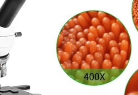
@micke thank you for sharing the videos. Those are not that bad actually, considering the hardware that you are using. One thing I should say (although you might already know), the pictures you see on Amazon are certainly not taken with the product advertised (whether it be the cameras or microscopes), so don’t expect that kind of quality. Also, most of the pictures they put are from sectioned samples. Like this one:
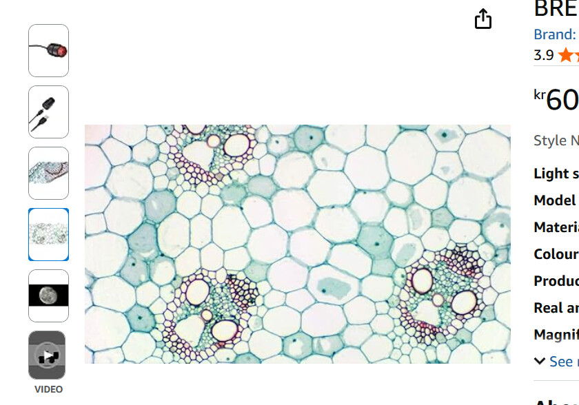
I am fairly sure this is a section that has been sliced down to 20-30um. If what you are imaging is thicker than 50um roughly, you’ll get a lot of blur from out-of-focus regions and it will reduce overall contrast.
This below is downright fake, it is possible to get such a 3D impression of an image with some microsocpes (such as DIC), but certainly not with the one advertised.
When you say you want to improve the quality of images and movies. Can you explain what you dislike in the videos you attached? Is it the graininess in the images, or you find it blurry?
Going to 4K is not a guarantee of better quality because the pixels tend to become smaller as well. As we’ve discussed, the pixels you have are already quite small for microscopy. For the same exposure time, smaller pixels will collect less light. There’s a whole discussion to be having about noise but if I try to put it simply:
- You have the read noise of your camera, this is a fixed amount of noise per image, the smaller your signal (or amount of light if you will) the more difficult it is to distinguish it from the read noise
- You have the shot noise of the light, this is a type of noise that is due to the discrete nature of light (photons), and scales with the square root of your amount of light. The relationship is not linear but having less light means your signal gets closer to the shot noise
One solution would be to increase the exposure time so that your smaller pixels collect the same amount of light. Yes, but:
- You have the thermal noise, this is a type of noise that scales (generally linearly) with the exposure time, it is heat that contributes to the false detection of a signal, and therefore increasing the exposure time leads to an increase in thermal noise
All cameras are different, and their noise levels vary quite a bit. But my main message is that having more pixels is not always a sign of quality. For reference, one of the standard camera we use in our microscopy facility are basically 2K with pixels that have a size of 6.5um.
Take care,
Omni