I have a Zeiss Axioobserver microscope, and a EMCCD camera, and wish to purchase a laser light source, and to set up a single molecule imaging system in my lab (I am a PhD student). However, I am ignorant of a good way to couple the lasers to the microscope.
In particular, I want to use the 100x, 1.4 numerical aperture objective, and to achieve a maximum power of 30 kW/cm^2 with a far-red laser of ~640nm, but to achieve much lower powers of 30 W/cm^2 with a 405nm, 488nm and 549nm laser. The illumination field should be at least 30 microns by 30 microns. I would like a Widefield, homogenous, square illumination field.
What do you recommend regarding laser engines and couplers, for a non-expert in optics, who is comfortable with reading the manual? A single component or very few components that I might purchase and easily assemble, in order to connect the high/low power lasers (uv,blue,red, and far-red), to the microscope, and to get Widefield illumination, would be ideal. Because the lab is an experimental biology lab, a coupling solution/part that couples the laser while completely encasing the laser beam is ideal.
I would be happy to hear also recommendations about which companies to consult with.
It sounds like you are interested in doing single molecule localization microscopy (SMLM)? If you haven’t already, probably the first person to ask is your Zeiss representative. They likely sell attachment(s) that would do what you are looking for and that would be easier to install and safe for most users.
Lumencor (https://lumencor.com/) is one company that sells laser engines who might be worth contacting.
I know the folks who created DNA PAINT had a super simple setup that included some laser pointers that they put together after the initial publication. Check out some of their papers–not the first, but something early.
Purely from optics point of view:
-if you illuminate the objective with a large collimated beam, you get a focused beam out
-on the contrary, if you want to get a (roughly) collimated beam out, you need to focus your laser in the back focal plane of the objective. This means for instance placing a 20 cm lens something like 19-20 cm from the objective. The illumination will not be homogenous, but will rather have a roughly Gaussian profile.If you want to do better than that, you’ll have to use additional tricks.
Moreover, if you now have separate lasers, you’ll need a beam combiner.
Goal: live‑cell HaloTag imaging (5 ms exposures) with a Gaussian spot whose central circular core is 30 µm in diameter and maintains ≥ 87 % of peak intensity, yielding 3 kW cm⁻² within that region.
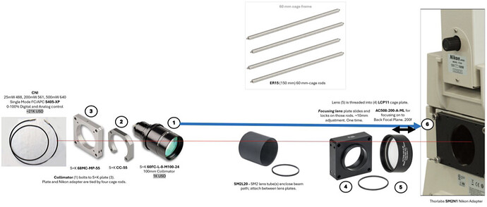
I will use, e.g., the Thorlabs Nikon Eclipse Ti Epi-Illumination Port Adapter (see image below) and lenses. After collimating the laser beam, and possibly also beam expanding it, do I need a lens to focus it onto the objective’s back focal plane (BFP), or does the Nikon Ti-E’s optical path inside the microscope body include lenses that perform this function?
In a previous response, @pawlowska suggested placing a 20 cm lens 19–20 cm from the objective—does this imply I must provide the lens that focuses on the BFP, and how can I confirm the exact distance from the Epi-Illumination Port at which to place the lens?
A Nikon Ti-E usually will not have a lens in the optical path between the filter and epi-illumination port. You could verify this by removing an objective and looking into the turret. If you can see an undistorted reflection of something like a cell phone light off of a dichroic in the filter wheel there is no lens in the optical path.
A 20cm lens could be a good choice, it depends on the diameter of your expanded and collimated input beam. The lens you choose and the objective will make a telescope with a demagnification proportional to the ratio of the focal lengths of the lens and the objective. If you use a 20cm lens and a 100x objective (approximately 2mm focal length for a Nikon) your expected beam size after the objective in the FOV is (input diameter) x 2mm/200mm. So if your input beam had a diameter of 10mm it would be 100um in the FOV. I think most people find it easier to use a longer focal length lens (so it is outside the body of the microscope) and a correspondingly large input beam diameter. Having the lens outside of the body also makes it easier to position.
How do you know you have the lens at the right distance? In my experience the easiest approach is to have the lens mounted on a rail or in optical cage so you can slide it back and forth. When the lens is at the right position the beam coming out of the objective is collimated. In practice you just adjust the lens position until the beam coming out of the objective is as small as possible at some point a good distance away from the microscope, like the ceiling of the lab. Then there is the X/Y alignment, for this it’s helpful to have the lens mounted on something like a Thorlabs CXY1A.